Team:KCL UK/Poster

Poster: KCL_UK


Poster Template

This poster template will let you create an interactive poster! The poster is divided in two parts: the visual overview on the left and the documentation on the right. The visual overview is broken down into sections that the user can click on. When a user clicks on a poster section in the visual overview, the documentation on the right will display the text and graphics associated with this section of the poster. You can find the documentation on how to use this template, as well as an example here: https://2020.igem.org/Competition/Deliverables/Poster

KCL iGEM 2020: Renervate

Project Title:

Creation of a 3D-bioprinted polycaprolactone scaffold with mussel-foot protein Pvfp-5β-based bioadhesive coating for biomedical applications.


Project Abstract:

Annually, between 250,000 and 500,000 individuals worldwide suffer from a spinal cord injury (SCI). SCI is characterised by damage to the spinal cord followed by a complex pathophysiological response and loss of neuronal function below the site of injury. The limited regenerative abilities of the CNS combined with the inhibitory environment created by the glial scar at the affected area pose numerous challenges to restoring function. Working towards a therapy for SCI, we have designed and modelled a biodegradable scaffold composed of polycaprolactone, that incorporates a synthetic mussel-foot protein based bioadhesive coating, to encourage axonal attachment, and can be produced using 3D-bioprinting methods. Our scaffold is customisable and contains the necessary micro- and macro-architectures predicted to topographically encourage axonal regrowth and withstand the mechanical forces in the spine. We have further investigated in silico the physicochemical properties of our chosen protein Pvfp-5β to better understand its biotherapeutic use.


Authors:

All authors are based at King's College London (London, United Kingdom) and are listed in alphabetical order.

Abigail Conner, Alya Masoud Abdelhafid, Ela Kanani, Emily Blundell, Gonzalo Leon Gonzalez, Harshraj Bumia, Ilaria Franceson, Jasmin Werner, Kyriakos Attouni, Kurt Peng, Leon Yong Kang Zhang, Liyamu Ma, Luke Bateman, Morgan Zvezdov, Peter Kanyike, Remy Tran, Shams Almusawi, Sonya Panchenko, and Stephanie Avraamides.


Sponsors:

We would like to express our sincerest thanks to Promega for their generous sponsorship. Additionally, we would like to thank King's College London for further funding. Thank you to SnapGene, Geneious, BMG Labtech, and The Biochemical Society for providing us with the software and financial assistance that has underpinned this project. We would also like to express our thanks to those who donated for our GoFundMe campaign.

Introduction: What are the aims of Renervate?

King’s College London iGEM 2020, or Renervate, is a group of 19 students working towards a novel treatment for spinal cord injury. Our overarching aim is to design and model a 3D-bioprintable nerve guidance channel/scaffold that contains a synthetic bioadhesive coating composed of a mussel foot protein, Pvfp-5β. The aim of this scaffold is to encourage axonal regeneration following traumatic injury and cell death in the spinal cord. Our project will be split into two phases.


The objectives of our Phase I research are outlined hereunder:

  1. Determine which scaffold macro-architecture is best for our aims (axonal regeneration) through Finite Element Analysis. Develop an algorithm using the visual coding language Grasshopper to provide the necessary scaffold micro-architectures for our aim.
  2. Develop a model that will predict how our scaffold will degrade in the body overtime.
  3. Provide a structural model for Pvfp-5β using GROMACS, and design an experimental procedure that can be used to create and validate the bioadhesive coating composed of this protein.

Inspiration:

Seeking inspiration from previous iGEM teams:

The inspiration for our project, Renervate, can be traced back to iGEM 2019. Our Team Leaders attended two pivotal presentations that would shape King’s College London’s 2020 project. Firstly, they attended Leiden’s presentation which revealed to them the potential of using protein-based hydrogels to treat burn wounds. Secondly, they attended the presentation by SCIE Great Bay whose project focused on categorising numerous mussel foot proteins (MFPs) with the intent of their use as bioadhesives. While watching these two presentations, our Team Leaders became inspired to look into the therapeutic applications of mussel-foot protein based biomaterials.

Biomaterial Scaffolds and Spinal Cord Injury: A Potential Cure?

Through their research, our Team Leaders became very interested in how this facet of biomaterials research can be applied to tissue engineering. In doing so, we found that we were able to incorporate our original ideas inspired by Leiden and Great Bay SCIE. Our preliminary research demonstrated to us that the main way in which biomaterials have been proposed as a treatment for SCI is as a bridging construct that facilitates axonal regrowth (Shrestha et al., 2014). The biomaterial “bridge” is referred to as a scaffolding and is used to restore tissue continuity in the injured area. Scaffolds intended to be used in SCI treatment can be split into two categories: hydrogels and nerve guidance channels.

The former are “hydrophilic, three-dimensional networks that are able to absorb large quantities of water or biological fluids” (quote from Chai et al., 2017). Hydrogels show great promise as they are injectable, flexible, and can be easily modified to contain elements that encourage nerve regrowth, such as biomolecule delivery and cell encapsulation (Straley et al., 2010). On the other hand, nerve guidance channels are channels that direct nerve growth from the proximal to distal nerve stumps and prevent regrowth into the scar. Similarly, nerve guidance channels can be designed to optimise regeneration by careful selection of macroarchitecture and microarchitecture features. With regards to SCI, hydrogels have been shown to be very promising as a consequence of their injectability, facilitating a much simpler implantation process in light of the complex geometries of the wound site. However, hydrogel technology still has far to come with regards to mechanical strength, degradation, cell-adhesivity, and topography. Therefore, our team decided to look into nerve guidance channel-based scaffolds, which have been shown to have the mechanical and physical properties required for implantation in the spinal cord.

Mussel Foot Protein Inspired Bioadhesives:

An essential element of scaffold design is adhesion. Adhesion is necessary to promote cell attachment to the scaffold as well as migration, proliferation, and differentiation. Mussel adhesive proteins (MAPs) have been the source of inspiration for numerous synthetic materials with biomedical applications, reviewed in the excellent paper by Kaushik et al., 2015. A previous study carried out by Murphy, Vollenweider, Xu and Lee in 2010 employed a MAP-inspired adhesive coating to a biologically inert scaffold for tendon repair. We were greatly inspired by this design and decided to look into how we could create a mussel foot protein-based bioadhesive polymer that coats the nerve guidance channel. The aim of the coating is to encourage attachment to the scaffold and therefore axonal regrowth along the bridge, eventually reconnecting the severved pathways.

Problem: Defining Spinal Cord Injury

How does spinal cord injury (SCI) affect the world?

Despite the severity of the clinical presentation of this condition, there is currently no cure for spinal cord injury. While there are numerous treatments available, they primarily focus on management and rehabilitation. In the United Kingdom, there are over 50,000 people currently living with SCI (Patek and Stewart, 2020), and in the US, a staggering 288,000 persons live with the condition (as of 2018). Beyond the physical effects, SCI places a tremendous burden on the economic and mental well-being of the individual. Nearly 48.5% of patients diagnosed with spinal cord injuries suffer from mental health problems, including depression, anxiety and PTSD (Migliorini et al., 2008). Additionally, the average lifetime cost of health and social care for a person with a spinal cord injury in the UK is £1.12 million (Mcdaid et al., 2019). In spite of the increase in access to care worldwide, the repercussions of SCI continue to pose a significant global challenge. Current treatments, such as immobilisation, surgical compression, and drug therapy do not focus on reconstructing axonal pathways severed by the injury. Therefore, they are not focused on restoring function but rather managing symptoms. Thus, we have decided to look into how Synthetic Biology and biomaterials can be used to provide a restorative solution to SCI.

What is the biological problem associated with SCI?:

At the biological level, SCI presents numerous challenges that must be overcome to provide a therapeutic solution. Firstly, the microenvironment of the spinal cord post-SCI becomes massively imbalanced due to the initial mechanical insult and biochemical tissue damage, which impairs functional recovery and axonal regeneration (Figure 1). Physical damage to the local capillaries of the spinal cord and severing of axons typically leads to the release of cytokines and chemokines into the extracellular space, depleting the blood-spinal cord barrier and increasing its permeability. The accumulation of macrophages in the microenvironment further promotes expression of cytokines and chemokines, as well as induces inflammation. Inflammation has a dual effect - it helps tissue remodelling and clear debris of lesion, but it also promotes further tissue breakdown (Fan et al., 2018). During the chronic phase of SCI, a glial scar forms which established both physical and chemical barriers to neuronal regeneration. Similarly, fluid-filled cysts can form at the site of injury and their extension during the chronic phase further contributes to cell death and axonal inhibition as well as the maturation of the glial scar. The glial cells serve to seal off the injury epicentre, secreting chemo repellents and growth-inhibiting matrix components (Alizadeh et al., 2019). We have devised a project design focussed on improving the function in the spinal cord post injury, through the resection of the glial scar and the implementation of our biocompatible scaffolding.

Figure 1: Illustration portraying the microenvironment and glial scar within the spinal canal as a result of SCI.

Our Idea:

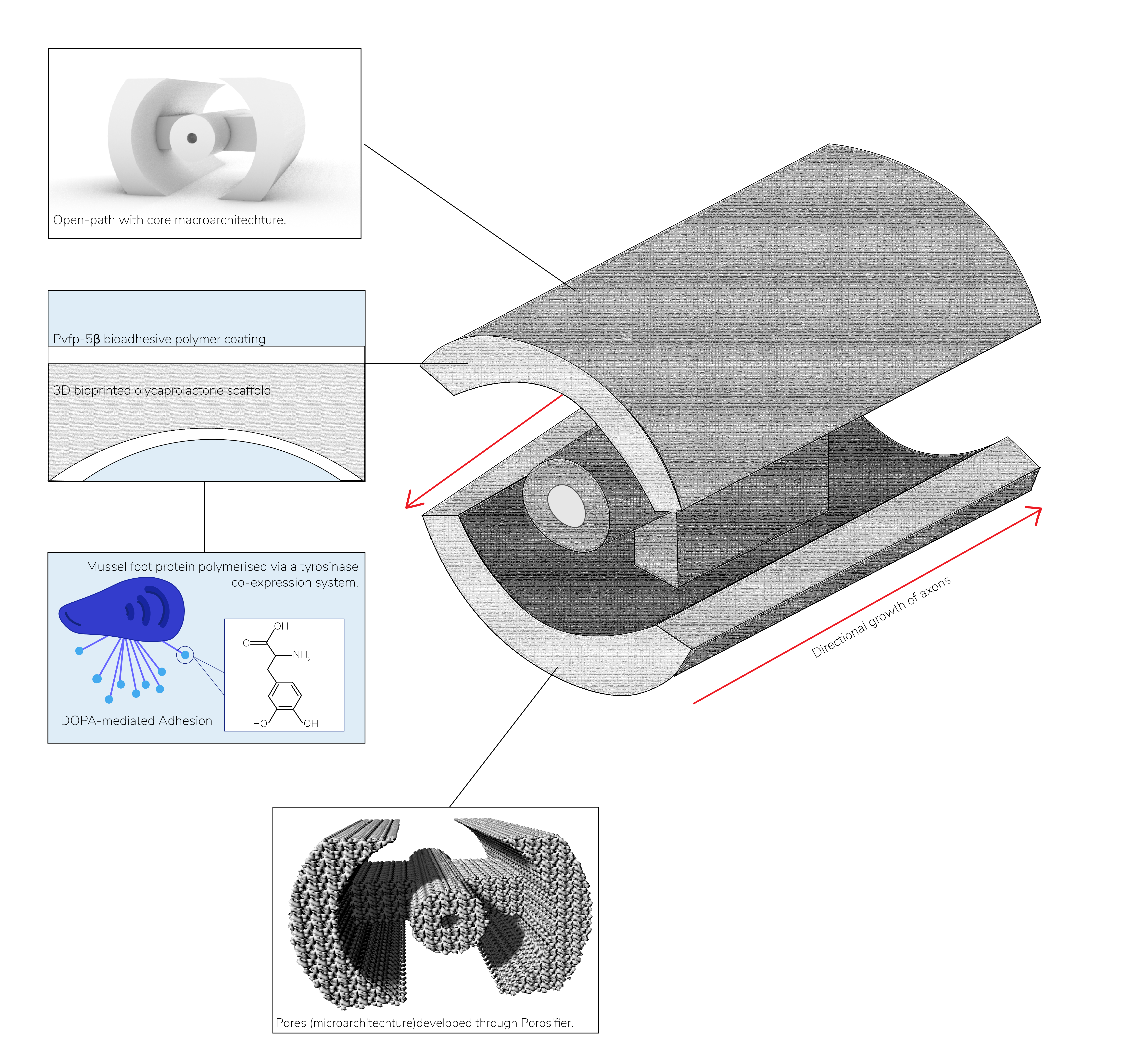
The Phase I project of Reneverate is focused on the design and modelling of a 3D-bioprinted polycaprolactone (PCL) based scaffolding that contains a novel mussel-foot protein based bioadhesive coating formed by Pvfp-5β. The second phase of our project will consist of the wet lab validation and formation of the scaffolding and the protein polymer. Our MFP coated scaffolding design has numerous potential applications in tissue engineering, yet we have designed it for use in treatment of SCI. The aim of our project is to outline a holistic, less invasive treatment that reduces the need for surgical intervention and focuses on functional restoration rather than management. To do so, we have designed an alternative method of glial scar removal prior to scaffold implementation and have carried out rational protein design to develop a novel, synthetic fusion protein that aims to encourage axonal regeneration in the spine. Furthermore, we have designed a biodegradable and biocompatible scaffolding that has been shown to withstand the mechanical forces of the spine and have modelled its degradation rate to ensure that it will remain in the spine for long enough to ensure healing without. Beyond the scientific aspects of our project, we have examined the societal role in SCI treatment. Consequently, Renervate has developed a Human Practices and Science Communication plan that ensures that we are able to tackle the social side of the problem.

Figure 1: Overview of our system. In the image above, our overall PCL-based scaffold design can be found. The open-path with core microarchitecture is evident in the overall structure as well as in the three-dimensional CAD model found in the top panel. Additionally, the use of a mussel-foot protein based bioadhesive polymer is visualised in the second panel from the top. The bioadhesive will coat the entire surface of the scaffold to provide an environment that encourages cell adhesion.

Our Engineering Design Cycle Success:

Spinal Cord Injury Engineering Success:

PCL Scaffold Design Engineering Success:

Mussel Foot Protein Engineering Success:

Scaffold Macro-architecture:

What is scaffold macro-architecture?

Different macro-architectures have been shown to have an influence on the success of scaffolds, with acellular scaffolds having a strong positive outcome when having a specific architecture (examples of such may be seen in Figure 1); ‘open-path’ designs allow better guidance with less material (as opposed to cylindrical designs) and permit the extension of nerve fibres across the entire defect length (Wong et al., 2008). A change in shape can lead to varying degrees of success when it comes to axonal regeneration, defect length and astrocyte migration. For example, the cylinder, tube and channel scaffolds had a doubling in the defect length, large scar and cyst formation and increased wall thickness of the prosthesis (the latter in the case of the tube scaffold). The two open path designs supported white matter tracts, allowed the extension of myelinated fibers and maintained the defect size over a period of 3 months with axonal regeneration observed.

One of the most important considerations for the macro-architecture of the scaffold are the mechanical properties. For example, the Young’s Modulus, or elasticity, of the scaffold should be as close to the spine as possible, and the scaffold should be able to withstand the forces in the environment without failure. We understood we needed to ensure our scaffold design took these into account, but there was a lack of prior studies regarding this design aspect. From Wong et al., 5 potential scaffold designs were evaluated using Finite Element Analysis in the Autodesk Inventor NASTRAN environment: cylinder, tube, channel, open path with core and the open path without core. This simulation allowed us to test for properties such as the von Mises stress, strain and displacement (Wong et al., 2008).

Figure 1: Macro-architecture designs implemented in Autodesk Inventor, based on the designs by Wong et al. (2008). (a) Open path without core, (b) Channel, (c) Tube, (d) Cylinder, (e) Open path with core

Our Final Design:

From our simulations none of the designs would have exceeded the yield strength (17.82 MPa) under a gravitational load, indicating that each one would have been viable. Therefore we compared the performance of the designs to each other and came to the conclusion that the open path with core was the best to use. It had the lowest stresses, strains and applied force. This result concurs with the literature review of Wong et al suggesting that this scaffold was also optimal for regrowth.

Scaffold Micro-architecture:

What is scaffold micro-architecture?

Micro-architecture is the surface structure of the scaffold. Alongside macro-structure, micro-structure is known to have a significant effect on cell behaviour and differentiation (Meco and Lampe, 2018). Subsequently, the axonal response to the scaffold is subject to vary dependent on the topography of the scaffold, as well as the size of the micro-structures (Zhang et al., 2020). Finally, our chosen material (polycaprolactone) does not possess suitable stiffness or cell adhesion properties in its dense form; following this, a solution to this issue outlined by Shahriari et al., is to introduce porosity - a commonly incorporated micro-architecture in scaffold engineering (Shahriari et al., 2017).

How have we incorporated micro-architecture in our design?

Following the decision to include pores in our scaffold design, we discovered that it was problematic to implement within our initially used CAD software, Autodesk Inventor. Therefore, we created an algorithm within the Rhinoceros Grasshopper 3D environment that has the capability of adding heterogeneous pores to any arbitrary shape - and so we made the software open-source. These added pores may be adjusted to modify the pore size and percentage porosity simply by altering the slider inputs for cell size and thickness. Utilising this script we created a final scaffold with an average pore size of 200µm and 58% porosity.

Figure 1: Original macro-architecture of scaffold (left) and porous scaffold design (right)

Scaffold Degradation Rate:

Why is the scaffold degradation rate important?

The length of time the scaffold needs to remain inside the body depends on the patient’s condition. The scaffold durability will need to be estimated through the simulation of its degradation inside the body to obtain a lifespan adaptable to the patient's needs.

Our Model:

After carrying out an extensive literature review, we created our own open-source MATLAB program to simulate the degradation of a polymer scaffold – as we could not identify a similar software to carry this out. Our program allows the user to find the prediction of degradation over time-based on a known degradation rate or scaffold porosity (given that the material is PCL for the latter). The calculation for degradation as a result of porosity is based on experimental data obtained by Zhang et al., 2013 specific to polycaprolactone(Zhang et al., 2013). However, if the user wishes to test another polymer scaffold’s degradation they may – if they know a specific degradation rate. There is the potential, nevertheless, for the code to be modified, such that the porosity mathematical relation is changed to fit another polymer. Moreover, it allows the user to enter the critical molecular weight (the limit of useful molecular weight) so that a warning dialogue can be displayed if their chosen design will decay too much in a specified time frame. If the initial molecular weight is unknown, there is also an option to find a suitable minimum initial molecular weight, providing that the duration that the scaffold must last, as well as a critical molecular weight and degradation rate/porosity, is given to the program. More information can be found on our GitHub.

Figure 1: User Interface for degradation program

Our Proposed Implementation:

We have organised the following workflow in order to synthesise Pvfp-5β in vivo, and undergo necessary experimentation and preparations for applications of our bioadhesive to a scaffold for therapeutic applications. This workflow is shown below in Figure 1. These steps will be completed in the near future, utilising our BioBrick assemblies and following our planned protocols as part of Phase II of our project, once we are able to gain access to laboratory equipment.

Figure 1: Our experimental workflow and proposed implementation with regard to our protein polymer.

We have assembled the following parts for our co-expression encompassing both Pvfp-5β (Figure 2a) and tyrosinase (Figure 2b) expression vectors. The sole component of our polymer is Pvfp-5β, a highly adhesive mussel foot protein, which can adhere to a variety of surfaces through different interactions (Lu et al., 2012), specifically via DOPA residues (Yu et al., 1999). We plan on polymerising monomeric Pvfp-5β using tyrosinase which catalyses the conversion of tyrosine residues into DOPA-quinone (Horsch et al. 2018), which readily undergo nucleophilic addition reactions between DOPA residues and cysteine residues of neighbouring DOPA residues (Ito and Prota, 1997). These reactions enable the formation of cysteinyl-DOPA bonds (in Figure 3) which are the crosslinks that hold our bioadhesive polymer together.

Figure 2: a) Pvfp-5β expression vector created through SnapGene. b) Tyrosinase expression vector created through SnapGene.

Our method of Pvfp-5β polymerisation is the subsequent method as a goal of improving overall bio-adhesive synthesis success, building on previous iGEM teams who also produced bioadhesives. Specifically, with our method of polymerisation, we aim to increase the yield of our adhesive, the main concern highlighted by Great Bay SCIE 2019.

Figure 3: Formation of cysteinyl-DOPA linkages from Tyrosinase - which results in polymerisation.

Following this, we will purify our bioadhesive polymer from our E.coli strain, and proceed to undergo various experimentation to analyse the suitability of our adhesive for applications to the scaffold, and the SCI site. After the approval of our product as a therapeutic device for treating spinal cord injury, our scaffold can be coated with our adhesive, ready for use in operations. We hope to successfully provide a novel treatment for individuals with SCI, as well as open pathways for future teams and researchers to explore further applications of our bioadhesive to biotechnology.

In preparation for unexpected results which do not meet standards, we have prepared mutagenesis studies with the hopes of improving our protein. This will be achieved primarily by using the R based software iGAM, developed by team Calgary 2019, resulting in the creation of a novel protein aimed at applications in SCI.

Structural Modelling of Pvfp-5β:

In order to better understand how Pvfp-5β adheres to various surfaces, we first needed to provide a working structural model to better understand which regions - and more importantly which residues - are involved in the adhesion mechanism (Figure 1). We decided to generate a homology model using the Phyre-2 fold recognition server (Kelley et al., 2015). Our Pvfp-5β target sequence from UniProt entry U5Y6U9, was used against a template model of Human Notch-1 EGFs 11-13 protein (PDB ID: 2VJ3). Our resulting homology model of Pvfp-5β was produced with a query coverage of 84% and an identity confidence of 99.7%. To further develop our model, and to elucidate one missing disulphide bond (C86-C95) - we employed Molecular Dynamics simulations using the GROMACS MD package (Berendsen, van der Spoel and van Drunen, 1995) as per the recommendation of our Instructors; Professor Annalisa Pastore and Dr Caterina Alfano. After a series of energy minimisation using GROMACS to bring down the distance between our desired residues, we used the YASARA Graphical modelling software (Krieger and Vriend, 2014) to impose a disulphide bond between residues C86 and C95. Our final model, with all known disulphide bonds is shown in Figure 2.

Figure 1: ‘Cartoon’ representation of Pvfp-5β shown in blue. All Tyrosine (Y) residues are shown in purple, as ‘sticks’. Tyrosine residues are inadvertently involved in the adhesion mechanism by their oxidation into 3,4-dihydroxyphenylalanine (DOPA).
Figure 2: 'Cartoon' depiction of Pvfp-5β (blue) model resulting from successive simulations using GROMACS, and YASARA. All (four) disulphide bonds are depicted as 'spheres'. From left to right; C25-C36, C48-C57, C86-95, C126-C135.

Education & Inclusivity Research:

Science Communication & Education:

To bring awareness to the applications of synthetic biology in a range of fields, we have spoken at virtual meet-ups due to the coronavirus pandemic. At these meet-ups we engaged with not only people who are aware of and interested in Synthetic Biology (as in our society’s introductory event), but also with audiences in which members come from different sectors- such as Policy Makers, Engineers, and Historians (such as in the Climate Tea Talk event). As our team has worked almost entirely virtually due to the coronavirus pandemic, we aimed to raise awareness on how synthetic biology may be used to combat the virus. In an effort to clarify misconceptions not only on the virus, but also Synthetic Biology, we created dialogue around different experiences and perspectives during the pandemic. We aimed to spread awareness through creating discussion videos, conveying scientific research through infographics, and participating in podcasts.

Our Biologix Competition & Inclusivity Research:

Aims and Overview of our Biologix Competition:

Additionally, we decided to investigate representation within the iGEM and the STEM community. Based on the obtained results from our research, we then aimed to highlight the existing barriers and decide on how to break them. A key area we identified has been the financial accessibility in iGEM and opportunities in the STEM field. In response to this, we designed an iGEM-influenced, innovative entrepreneurial competition, Biologix. This is a free biotechnology and synthetic biology competition, giving an amazing opportunity for all penultimate year high schoolers to take part in a project. Through Biologix, we aim to give students from different school backgrounds, the opportunity to participate in an outstanding project during their high school career, which is made inclusive as it will be solely based on literature research avoiding discrimination towards schools not provided of laboratory facilities and/or enough funds for equipment. The Biologix mission is to give students an accessible opportunity to develop a passion for synthetic biology, encouraging them to pursue a career in the field. Providing assistance with university application and interviews, and inspiring students to start partaking in university iGEM teams.

Further Research into Intersectionality & STEM:

Furthermore, as previously stated, we also focused on the STEM field in general, analysing which are the existing barriers and how we could overcome them. Our focal point has been the United Kingdom as this represents our local community. From the information collected we discovered that teachers are underfunded as well as having minimal training in STEM subjects; additionally, the effect of social beliefs on enforcing gender gaps and exclusion of minorities has been highlighted.

Finally, to assess the previously discussed matters we provided an “Intersectionality in STEM” guide, which can be accessed on the Inclusivity page, in which we proposed some strategies to be implemented in the STEM community to support minorities and combat discrimination.

Entrepreneurship:

Our product can fill a gap in the market due to the lack of treatments for patients with syringomyelia in the cervical thoracic spine. We identified several enterprises developing products within the same niche market, but the uniqueness of our scaffold falls on the use of our synthetic MFP based protein which makes Renervate stand out. To be able to put our product on the market there is a procedure to be followed depending on the intended market, in our case our targeted market is the EU, where the first step is to obtain a patent for our scaffold through the European Patent Office. Next step is to make sure our device falls within the legislative frame of the CE marking for medical devices settled by the Medical Device Regulation, published in 2017 by the European parliament (Johner Institute, 2020). To conclude, we have established a three-step strategy which we designed with Dr Prashant Jha’s guidance, to obtain the CE marking:

  • To submit a request for a CE certification of an experimental structure
  • To carry out clinical trials
  • To apply for CE certification as a Medical Device
  • The centrepiece in the application for the CE marking falls on the clinical trials that must prove its safety and correct functioning. We have organised trials in 6 phases with the advice of Dr Prashant Jha, described in the following diagram:

    The literature review as well as part of the modelling have already been carried out and we intend on continuing modelling and begin in vitro studies during phase 2 of Renervate. Our timeline predicts clinical investigations could last between 5 and 10 years.

    Mitigating safety hazards is highly important to put a product in the market, even more for a class III device like ours. Therefore, we investigated pre-existing hazards of similar devices reported on the MAUDE database and found 5 issues to be considered in our clinical trials:

    • Medical malpractice
    • Nerve damage due to the device
    • Detachment of site of injury
    • Infection
    • Interaction with nearby tissue

    We also informed ourselves about the procedure for the FDA due to its strong recognition worldwide.

References:

  1. Alizadeh, A., Dyck, S.M., Karimi-Abdolrezaee, S., 2019. Traumatic Spinal Cord Injury: An Overview of Pathophysiology, Models and Acute Injury Mechanisms. Front. Neurol. 10, 282. https://doi.org/10.3389/fneur.2019.00282
  2. Berendsen, H., van der Spoel, D. and van Drunen, R., 1995. GROMACS: A message-passing parallel molecular dynamics implementation. Computer Physics Communications, 91(1-3), pp.43-56.
  3. Fan, B., Wei, Z., Yao, X., Shi, G., Cheng, X., Zhou, X., Zhou, H., Ning, G., Kong, X., Feng, S., 2018. Microenvironment Imbalance of Spinal Cord Injury. Cell Transplant 27, 853–866. https://doi.org/10.1177/0963689718755778
  4. Haynes, L., 2008. Studying Stem? What Are The Barriers?. [online] Mei.org.uk. Available at
  5. Horsch, J., Wilke, P., Pretzler, M., Seuss, M., Melnyk, I., Remmler, D., Fery, A., Rompel, A., & Börner, H. G. (2018). Polymerizing Like Mussels Do: Toward Synthetic Mussel Foot Proteins and Resistant Glues. Angewandte Chemie (International ed. in English), 57(48), 15728–15732. https://doi.org/10.1002/anie.201809587
  6. Ito, S., & Prota, G. (1977). A facile one-step synthesis of cysteinyldopas using mushroom tyrosinase. Experientia, 33(8), 1118–1119. https://doi.org/10.1007/BF01946005
  7. Johner Institute, “Starter-Kit: How To Quickly, Easily And Safely Master The Medical Device Approval Process” (https://www.johner-institute.com/starter-kit/) accessed October 2020.
  8. Kaushik, N., Kaushik, N., Pardeshi, S., Sharma, J., Lee, S. and Choi, E., 2015. Biomedical and Clinical Importance of Mussel-Inspired Polymers and Materials. Marine Drugs, 13(11), pp.6792-6817.
  9. Kelley, L., Mezulis, S., Yates, C., Wass, M. and Sternberg, M., 2015. The Phyre2 web portal for protein modeling, prediction and analysis. Nature Protocols, 10(6), pp.845-858.
  10. Krieger, E. and Vriend, G., 2014. YASARA View—molecular graphics for all devices—from smartphones to workstations. Bioinformatics, 30(20), pp.2981-2982.
  11. Lu, Q., Danner, E., Waite, J.H., Israelchvili, J.N, Zeng, H. and Hwang, D.S., (2012). Adhesion of mussel foot proteins to different substrate surfaces. J R Soc Interface 10: 20120759.
  12. Mcdaid, D., Park, A.-L., Wahlbeck, K., 2019. The Economic Case for the Prevention of Mental Illness. Annu. Rev. Public Health 40.
  13. Meco, E. and Lampe, K. J. (2018) ‘Microscale Architecture in Biomaterial Scaffolds for Spatial Control of Neural Cell Behavior’, Frontiers in Materials, 5, p. 2. doi: 10.3389/fmats.2018.00002.
  14. Migliorini, C., Tonge, B. & Taleporos, G., 2008. Spinal Cord Injury and Mental Health. Australian & New Zealand Journal of Psychiatry, 42(4), pp.309–314.
  15. Morgan, R., Kirby, C. and Stamenkovic, A., 2016. The UK STEM Education Landscape. [online] Raeng.org.uk. Available at: https://www.raeng.org.uk/publications/reports/uk-stem-education-landscape
  16. Murphy, J., Vollenweider, L., Xu, F. and Lee, B., 2010. Adhesive Performance of Biomimetic Adhesive-Coated Biologic Scaffolds. Biomacromolecules, 11(11), pp.2976-2984.
  17. Patek, M., Stewart, M., 2020. Spinal cord injury. Anaesth. Intensive Care Med. 21, 411–416. https://doi.org/10.1016/j.mpaic.2020.05.006
  18. Shahriari, D. et al. (2017) ‘Hierarchically Ordered Porous and High-Volume Polycaprolactone Microchannel Scaffolds Enhanced Axon Growth in Transected Spinal Cords’, Tissue Engineering Part A, 23(9–10), pp. 415–425. doi: 10.1089/ten.tea.2016.0378.
  19. Wong, D. Y., Leveque, J.-C., Bumblay, H., Krebsbach, P. H., Hollister, S. J., & LaMarca, F. (2008). Macro-Architectures in Spinal Cord Scaffold Implants Influence Regeneration. Journal of Neurotrauma, 1027-1037.
  20. Yu, M., Hwang, J. and Deming J.T., (1999). Role of l-3,4-Dihydroxyphenylalanine in Mussel Adhesive Proteins J. Am. Chem. Soc. 121(24), 5825–5826.
  21. Zhang, D. et al. (2020) ‘Physical understanding of axonal growth patterns on grooved substrates: groove ridge crossing versus longitudinal alignment’, Bio-Design and Manufacturing, 3(4), pp. 348–360. doi: 10.1007/s42242-020-00089-1.
  22. Zhang, Q., Jiang, Y., Zhang, Y., Ye, Z., Tan, W. and Lang, M., 2013. Effect of porosity on long-term degradation of poly (ε-caprolactone) scaffolds and their cellular response. Polymer Degradation and Stability, 98(1), pp.209-218