Contribution
This page has the same content with the page Measurement
Overview
To make a useful contribution for future iGEM teams. In this year, we constructed an EGFP expression vector (with pSB1C3 as the plasmid skeleton) in which the expression of EGFP was controlled by the promoter BBa_K1402010 and transformed it into E.coli BL21 (DE3) which is commonly used to express protein. Then, by measuring the EGFP fluorescence, we investigate the optimal IPTG concentration and temperature for the expression of protein in E.coli BL21 (DE3) using the BBa_K1402010 in pSB1C3. Meanwhile, we carried out RT-qPCR to directly investigate the relation between the original strength of BBa_K1402010 and IPTG concentration at the transcription level without interfering by context.
Vector Construction
Our part vectors contained EGFP were successfully constructed and sequence results were correct. With the induction of IPTG, as shown in Figure 1, we can see the green fluorescence obviously under the stimulate of blue light.

Figure 1. Fluorescence of EGFP after the promoter BBa_K1402010 with the induction of IPTG.
Fluorescence Result
Then we picked the positive clone in Figure 1 for further experiment. When the E.coli BL21 (DE3) was cultured at the OD600 between 0.6 to 0.8, we added IPTG in different concentration and cultured the E.coli in different temperature. Absolute fluorescence and OD600 assessed by fluorescence microplate reader was shown below.


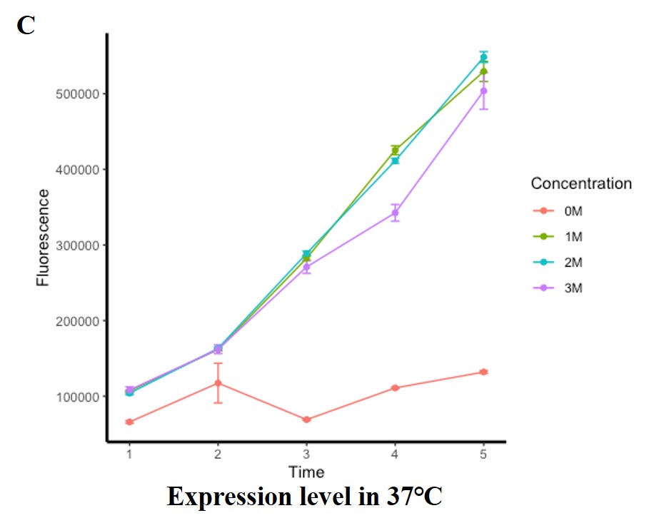
Figure 2. Fluorescence of EGFP under different treatment condition. (A) (B) (C) represents the treatment at 25℃, 30℃ and 37℃ respectively. The horizontal axis shows the different concentrations of IPTG, the vertical axis shows the green fluorescence per OD600 (excitation wavelength: 485 nm; detection wavelength: 528 nm), and segments and data points of different colors show the different culture temperature. Error bar indicates the standard error of replicates.
As shown in Figure 2 above, we can find that even through without induction of IPTG, fluorescence can be measured, suggested that when BBa_K1402010 was used to express protein in E.coli BL21 (DE3) with pSB1C3 as the plasmid skeleton, it is leaky. But the leaky expression is unstable and it is slower than the degradation rate in some condition as shown in Figure 2A red line.
Compared the expression level in different temperature, fluorescence in 37℃ was much higher than in 25℃ and 30℃, indicating that the optimum temperature for BBa_K1402010 expressed in BL21 with pSB1C3 as the skeleton is 37℃. And the optimum IPTG concentration in 37℃ was 2 mM.
RT-qPCR Result
From the results of fluorescence characterization, 37℃ seems to be a more favorable temperature for the opening of BBa_K1402010 in pSB1C3 in E.coli BL21 (DE3), so we chose this temperature as the culture temperature for RT-qPCR. We selected a frequently used housekeeping genes 16srRNA as reference genes to compare the relative expression level of EGFP controlled by BBa_K1402010 under different IPTG concentrations of 1, 1.5, 2, 2.5 and 3 mM at 37℃.

Figure 3. The relative normalized expression level of EGFP of E.coli BL21 (DE3) under the conditions of IPTG concentration of 1, 1.5, 2, 2.5 and 3 mM at 37℃. The horizontal axis shows the different concentrations of IPTG, the vertical axis shows the relative normalized expression level of EGFP of each test group.
From Figure 3, we can conclude that the optimum IPTG concentration is 2.5 mM, which is corresponding to the result in Figure 2C. However, the expression level between 1 mM and 2 mM IPTG showed a slow downward trend. We speculated that the actual expression level from 1 mM to 2 mM IPTG were very closed due to both of them were not the most suitable for BBa_K1402010, but the degradation rate caused by different bacterial denisity were different, resulted in a slow downward trend.
Protocol
Fluorescence Characterization
1. Linearize pSB1c3 plasmid by PCR.
(F: TACTAGTAGCGGCCGCTGCAG, R: CTCTAGAAGCGGCCGCGAATTC)
2. Obtain the sequence of BBa_K1402010 from 2019 DNA Distribution Kit by PCR.
(F: GAATTCGCGGCCGCTTCTAGAG, R: CTGCAGCGGCCGCTACTAGTA)
3. Obtain the RBS+EGFP sequence and part sequence by PCR. Plasmid contained EGFP sequence was
kindly donated by Associated Prof. Wentao Jin, sequence was identical to UniProtKB -
A0A2V2QJP9.
(F: AAAGAGGAGAAATACTAGATGAGCAAGGGC, R: TTACTTGTACAGCTCGTCCATG)
4. Use homologous recombination to construct the BBa_K1402010-EGFP pasmid.
5. Transform the recombination plasmids into E.coli
DH5α, then pick the positive clones and sequence.
6. After recombinational sequence results were comfirmed, transform the plasmids into E.coli BL21 (DE3).
7. Coated the transformed BL21 onto the plate and added 1 mM IPTG.
8. Incubated at 37℃ overnight and observed colony color under the blue light to see if the EGFP
is expressed.
9. Picked greener single colonies, add them into 1 mL LB, incubate at 37℃ in a shaker for 6–8 h
till the OD600 reach 0.6–0.8.
10. Add 50 μl germ solution from last step into 5 mL LB, add IPTG in different concentration and
induce for 5 h at 25℃, 30℃ and 37℃.
11. Measure the fluorescence (excitation wavelength: 485 nm; detection wavelength: 528 nm) and
OD600.
Fluorescence Characterization
1. Pick a single colony from BBa_ K1402010 and inoculate in 5 mL liquid LB medium + 5 μL
Chloramphenicol (25 mg/mL in EtOH) and grow the cells overnight at 37°C and 220 rpm.
2. Inoculate 50 μL of the overnight culture into 4.95 mL LB with Chloramphenicol to make 7
parallel groups and grow the cells at 37°C and 200 rpm until OD600 reaches 0.6.
3. Add IPTG to final concentrations of 1, 1.5, 2, 2.5 and 3 mM IPTG respectively and
grow the cells at temperature of 37°C and 200 rpm for 5 hours.
4. Extracted RNA, make reverse transcription and carry out RT-qPCR with 16srRNA as reference
genes. RT-qPCR data was proceeded by Bio-Rad CFX Maestro.
RT-qPCR primer for EGFP:
F: AGATCCGCCACAACATCGAG, R: AACTCCAGCAGGACCATGTG
RT-qPCR primer for 16SrRNA:
F: AACACATGCAAGTCGAACGG, R: TAAGGTCCCCCTCTTTGTGC

