Team:ZJU-China/Engineering

Engineering
Overview
Our project is typically problem-based. And each step to support engineering success follows the design cycle as iGEM suggests, which starts from researching, and is followed with imagination, designing, building, testing, learning, and improving, and continues with a re-research again. Thankfully, with the effort made by every member of ZJU-2020, we constructed two pivotal parts of our project to demonstrate our design was workable by both wet experiments and modeling analysis. And the whole process of our engineering is as below.
Research
As we mentioned in the background of our project, there are obvious disadvantages of present contrast agents for MRI of not only breast cancer but all kinds. We wanted to make improvements in the specificity of contrast agents, and we understood clearly that we must create a modified contrast agent to meet the standard of specificity. Adding targeting molecule on gadolinium, which is now widely used for MRI was first proposed but quickly dismissed for the toxicity of gadolinium. And then we started to search if there is a biocompatible magnetic material for MRI, and we finally found a biological one, which is called magnetosome as our product.
For the proper choice for targeting a molecule, the antibody was aforethought. We also studied other kinds of molecules that targeted HER2 cells such as peptides which tended to the acidic environment of tumors; nucleic acid aptamers that bind HER2 specifically and so on. For the convenience of design and building a standardized platform in the future, we finally chose a single-chain variable fragment (scFv) as the targeting molecule for HER2.
Imagine & Design
By studying the protocols of the transgene on MSR-1, we can obtain the surface-modified magnetosomes by transferring the recombinant plasmid containing the fusion protein of our wanted protein and mamC (membrane protein covers the most area of the surface of magnetosomes) into magnetotactic bacteria, using biparental conjugation. So at first, we designed to link scFv directly to mamC to functionalized the magnetosomes.
However, no research showed such a design like us. Instead, they preferred to choose a third one as an anchor for the molecule that they wanted to link to mamC in case that the magnetosomes would be unfunctional for direct linkage. Refer to those researches, we decided on ZZ, the Fc-binding domain of an immunoglobulin-binding protein from the cell wall of Staphylococcus aureus as the anchor and add Fc to scFv for fusion with mamC-ZZ (Figure 1).
For more detailed information about our design, you can visit the Design page of our wikis.

Figure 1. (A) Map of mamC-ZZ vector; (B) Map of scFv-Fc vector.
Build
The building process is described as the experiment part of our wikis, we constructed mamC-ZZ and scFv-Fc proteins as we designed according to recent studies. And we submitted them to the iGEM Registry following the BioBricks assembly standard.
For more detailed information about our parts, we strongly recommend you visit the corresponding parts pages on the iGEM BioBricks Registry.
Testing
In the wet experiments, we first confirmed the sequences of the two fusion proteins we designed by amplifying them from PCR (Figure 2). And the sequencing results showed no difference with the maps of the two vectors.
For more detailed information about our experiments, you can visit the Results and Model part of our wikis.

Figure 2. (A) scFv-Fc amplified from pET19b; (B) mamC-ZZ amplified from pGEX-2TK.
Then, we transferred pET19b_scFv-ZZ and pGEX-2TK_mamC-ZZ into E.coli SHuffle® and BL21 (DE3) to express the fusion proteins respectively and obtained the purified proteins successfully (Figure 3).

Figure 3. (A) Western blotting results of immunoprecipitation of scFv-Fc; (B)&(C) Western-blot results of GST mamC-ZZ under different inducing IPTG (B) concentration and inducing time (C).
To validate the interaction between mamC-ZZ and scFv-Fc, co-immunoprecipitation was carried out and the results turned out that the two fusion proteins bound together specifically (Figure 4).

Figure 4. Western blot results of mamC-ZZ and scFv which introduce different primary antibody.
In order to prove that scFv-Fc fusion protein can specifically target HER2-positive breast cancer cells, we have demonstrated the specificity and effectiveness of this targeting by flow cytometry (Figure 5).

Figure 5. Flow cytometry results of MDA-MB-453 and MDA-MB-231 after incubated with scFv-Fc.
As MSR-1 was microaerophilic, we added resazurin into the liquid medium to see the growth of cells by the change of the color. The results showed that the color of the liquid medium which was added into MSR-1 was obviously different from that without the addition of MSR-1 (Figure 6). To further prove that the magnetotactic bacteria were truly cultured, bacterial PCR was carried out using the primers designed from the 16srDNA of Magnetospirillum magneticum and we used E.coli and Agrobacterium as control. The results showed that the magnetotactic bacteria were indeed grown (Figure 7).

Figure 6. Liquid medium cultured MSR-1 (red test tubes) contrast to the blank control ( blue test tubes).

Figure 7. Results of the bacterial PCR.
In the modeling analysis, we mainly focused on three aspects of our product, including the yield of scFv-Fc and magnetosomes in their expression chassis, the combination and disaggregation of these two proteins and the interaction of HER2 and the modified magnetosomes in vivo.
In the first part, we constructed two deterministic models to compute the production of scFv-Fc and modified magnetosomes. And the results showed that the expression of these two proteins can be precisely and highly activated after the addition of IPTG (Figure 8,9).

Figure 8. Dynamics of target protein. Horizontal axis shows the length of time. Vertical axis demonstrates the amount of protein (scFv-Fc) within the system.

Figure 9. Dynamics of target protein. Horizontal axis shows the length of time. Vertical axis demonstrates the amount of protein (mamC-ZZ) within the system.
In the second part, we constructed a deterministic model to determine the combination and disaggregation of scFv-Fc and modified magnetosomes in vitro and it showed that they bound rapidly in a very short time, ensuring that the feasibility of auto-assembling in vitro (Figure 10).
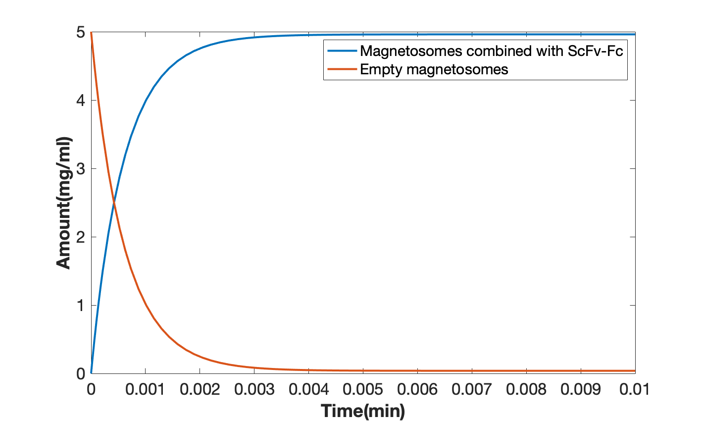
Figure 10. Combination of scFv-Fc and modified magnetosomes (the blue line refers to the combination product of scFv-Fc and modified magnetosomes, and the orange line refers to pure magnetosomes).
In the last part, we constructed a model about modified magnetosomes in vivo to describe the combination and degradation with HER2 and the complex of our modified magnetosomes and scFv-Fc. It turned out that the complex can bind HER2 rapidly while degraded in a relatively long period of time (Figure 11,12), which makes it potential for rapid detection and long-term treatment if we develop it into a drug delivery vector.

Figure 11. Magnetosome binding in a short time.

Figure 12. Metabolism of magnetosomes in the body for a long time.
For more detailed information about our modeling, you can visit the Model page of our wikis.
Actions under COVID-19
Due to the spread of the COVID-19 virus, the majority of our lab work has been delayed. For example, because MSR-1 was not a common strain like E.coli, we need to purchase it from abroad to obtain quality strains. Besides that, the cultural condition had a tough standard which should be taught by experienced technicians. Though we connect a professor studying the culture of magnetotactic bacteria, yet teaching the methods simply through the internet is basically useless for there being so many details culturing this microaerobic bacteria. However, for making some progress in the area of contrast agents, we still tried our best to carry out relative experiments to demonstrate our design and listed the plans for the following experiments. For the detailed content of the future work, we set a wiki page specialized for it.
